LabAdviser/314/Microscopy 314-307/TEM/ATEM: Difference between revisions
mNo edit summary |
|||
| (8 intermediate revisions by one other user not shown) | |||
| Line 4: | Line 4: | ||
[[Category:314]] | [[Category:314]] | ||
[[Category:314-Microscopy]] | [[Category:314-Microscopy]] | ||
''This section is written by DTU Nanolab internal if nothing else is stated.'' | |||
[[File:Warning Computer Machine.png|left|100x100px]] | |||
== The Titan ATEM is not available at DTU Nanolab any longer. The information here is just for reference. == | |||
<br /> | |||
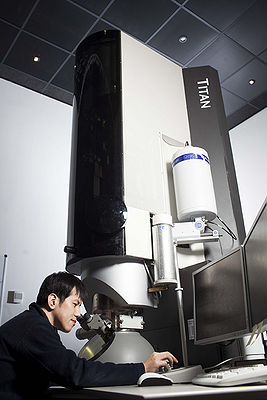
[[image:8212.JPG|400x400px|right|thumb|Titan ATEM in building 314.]] | [[image:8212.JPG|400x400px|right|thumb|Titan ATEM in building 314.]] | ||
| Line 11: | Line 18: | ||
Titan Analytical can be used in two conditions, transmission (TEM) and scanning transmission (STEM) modes. It is equipped with the FEI X-FEG gun and monochromator, which allows experiments at 120 kV or 300 kV acceleration voltage. With the momochromator, the energy spread can be adjusted between about 1 eV and below 0.2 eV. Furthermore, a CEOS CESCOR Cs-corrector of the condenser lens system is installed. The objective lens has the super-twin (S-Twin) pole piece with 5.2 mm pole gap (Cs about 1.2 mm). The point (interpretable) resolutions for TEM and STEM at 300 kV are 0.2 and 0.08 nm, respectively, which allows atomic arrangements in materials to be visualized clearly. For TEM, a Gatan Ultrascan US1000 CCD (2048x2048 px) is installed, for STEM, a high-angle annular dark field detector (HAADF) or BF/DF detector of the GIF system can be used.<br /> | Titan Analytical can be used in two conditions, transmission (TEM) and scanning transmission (STEM) modes. It is equipped with the FEI X-FEG gun and monochromator, which allows experiments at 120 kV or 300 kV acceleration voltage. With the momochromator, the energy spread can be adjusted between about 1 eV and below 0.2 eV. Furthermore, a CEOS CESCOR Cs-corrector of the condenser lens system is installed. The objective lens has the super-twin (S-Twin) pole piece with 5.2 mm pole gap (Cs about 1.2 mm). The point (interpretable) resolutions for TEM and STEM at 300 kV are 0.2 and 0.08 nm, respectively, which allows atomic arrangements in materials to be visualized clearly. For TEM, a Gatan Ultrascan US1000 CCD (2048x2048 px) is installed, for STEM, a high-angle annular dark field detector (HAADF) or BF/DF detector of the GIF system can be used.<br /> | ||
For energy dispersive X-ray spectroscopy (EDS) an Oxford windowless X-Max | For energy dispersive X-ray spectroscopy (EDS) an Oxford windowless X-Max 100TLE detector is installed, which is running Oxford Aztec analysis software. For electron energy loss spectroscopy (EELS) and energy filtered imaging (EF-TEM), the microscope is equipped with a Gatan Tridiem 865 GIF and a Gatan Ultrascan US1000 CCD (2048x2048 px). The microscope can be used for elemental analysis from regions that are as small as 1 nm and is specifically suited for EDS and EELS mapping (Gatan Digiscan). Specially, monochromated EELS, which is reachable to an energy resolution of 0.15 eV, allows the distribution of surface plasmons in nanostructured materials to be imaged at the nanometer scale and makes possible to determine the valence state of elements (e.g., Fe2+/Fe3+ ratios).<br /> | ||
This microscope is also dedicated to magnetic and electrostatic potential imaging since it has a biprism located at a selected-area aperture position and a Lorentz lens. This capability not only offers us to characterize magnetic materials and semiconductor devices but also may make possible to visualize different chemical states in low-density materials such as polymers and biological specimens. <br /> | This microscope is also dedicated to magnetic and electrostatic potential imaging since it has a biprism located at a selected-area aperture position and a Lorentz lens. This capability not only offers us to characterize magnetic materials and semiconductor devices but also may make possible to visualize different chemical states in low-density materials such as polymers and biological specimens. <br /> | ||
| Line 37: | Line 44: | ||
= Calibration = | = Calibration = | ||
*[[ | *[[Media:ATEM collection-angles.xlsx|GIF collection angles (20220209)]] | ||
*Magnification calibration cameras | *Magnification calibration cameras | ||
*Convergence angles STEM | *Convergence angles STEM | ||
= Process information = | = Process information = | ||
Latest revision as of 22:48, 16 June 2025
Feedback to this page: click here
This section is written by DTU Nanolab internal if nothing else is stated.

The Titan ATEM is not available at DTU Nanolab any longer. The information here is just for reference.

FEI Titan 80-300 ATEM
Titan Analytical can be used in two conditions, transmission (TEM) and scanning transmission (STEM) modes. It is equipped with the FEI X-FEG gun and monochromator, which allows experiments at 120 kV or 300 kV acceleration voltage. With the momochromator, the energy spread can be adjusted between about 1 eV and below 0.2 eV. Furthermore, a CEOS CESCOR Cs-corrector of the condenser lens system is installed. The objective lens has the super-twin (S-Twin) pole piece with 5.2 mm pole gap (Cs about 1.2 mm). The point (interpretable) resolutions for TEM and STEM at 300 kV are 0.2 and 0.08 nm, respectively, which allows atomic arrangements in materials to be visualized clearly. For TEM, a Gatan Ultrascan US1000 CCD (2048x2048 px) is installed, for STEM, a high-angle annular dark field detector (HAADF) or BF/DF detector of the GIF system can be used.
For energy dispersive X-ray spectroscopy (EDS) an Oxford windowless X-Max 100TLE detector is installed, which is running Oxford Aztec analysis software. For electron energy loss spectroscopy (EELS) and energy filtered imaging (EF-TEM), the microscope is equipped with a Gatan Tridiem 865 GIF and a Gatan Ultrascan US1000 CCD (2048x2048 px). The microscope can be used for elemental analysis from regions that are as small as 1 nm and is specifically suited for EDS and EELS mapping (Gatan Digiscan). Specially, monochromated EELS, which is reachable to an energy resolution of 0.15 eV, allows the distribution of surface plasmons in nanostructured materials to be imaged at the nanometer scale and makes possible to determine the valence state of elements (e.g., Fe2+/Fe3+ ratios).
This microscope is also dedicated to magnetic and electrostatic potential imaging since it has a biprism located at a selected-area aperture position and a Lorentz lens. This capability not only offers us to characterize magnetic materials and semiconductor devices but also may make possible to visualize different chemical states in low-density materials such as polymers and biological specimens.
Sample holders
The default specimen holders are a Fischione single-tilt tomography holder and an FEI double-tilt holder, which should be in the pumping station. The lab has also other specimen holders used for various application e.g. heating (furnace and MEMS-based), cooling, biasing and tomography. For information on the various specimen holders see HERE
Who may operate the Titan ATEM
In order to start training on the Titan ATEM you must by fully trained in the Tecnai TEM. Most of the basic operation on the Titan ATEM is the same as on the Tecnai with the added complexity of the aberration corrector and the monochromator.
When you have demonstrated a high level of familiarity with the microscope you are allowed to book it via LabManager and use it 24/7.
Booking
Booking on the ATEM is done by the users in accordance to the booking rules. Booking rules can be found in LabManager under "Documents" for the ATEM.
Further information
Calibration
- GIF collection angles (20220209)
- Magnification calibration cameras
- Convergence angles STEM
Process information
The following techniques and processes are available on the microscope (list isn't complete):
Techniques:
Processes:
- Monochromated STEM-EELS for low-loss experiments
Tips and Tricks
Here you can find some help to solve small issues on the microscope. Let us know, if you have other suggestions.
- Magic Switch is not working properly
- Aztec connection problems
- GIF apertures are not responding
- HAADF detector doesn't give a signal
- No buttons for STEM acquisition
Reference material
L. Reimer, Transmission Electron Microscopy - Physics of image formation and microanalysis (Springer, 1997).
David B. Williams, C. Barry Carter, Transmission Electron Microscopy - A Textbook for Materials Science (Springer, 2009).
