Specific Process Knowledge/Lithography/EBeamLithography/RaithElphyManual
Feedback to this page: click here
= THIS PAGE IS UNDER CONSTRUCTION
Purpose, location and technical specifications
OBSOLETE! This tool does not exist at DTU Nanolab any longer.

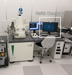
The Raith Elphy system is a pattern generator built onto the LEO Scanning Electron Microscope (SEM) in cleanroom F-2. All users must therefore acquire license to use the SEM LEO before acquiring license to the Raith Elphy system.
Remember that you need two additional trainings from Nanolab staff in order to be able to use this tool for lithography. Please refer to the General Page for further information.
The Raith ELPHY Quantum software (from now on, Elphy) is installed on the Raith pc located right next to the LEO SEM pc. The workstation is more or less independent from the LEO itself. If you try to access Elphy without properly setting up the microscope first, it will usually return a communication error, but it would still be partially functional e.g. to prepare layouts and position lists. When you want to do actual patterning, you will need to operate on both computers at the same time.
The pc has limited access to internet and the network. To import and export files on the Raith pc, two options are available:
- Send the files to yourself on your DTU email. You can then access and download them from the DTU webmail page
- For more advanced functionality, use the Citrix page with your DTU login. You can then use a Remote Desktop (DTU Office 2013 IE11 Desktop) or an App (DCH-LAB Windows Explorer) to operate and transfer files between the local pc and your personal M: drive.
Preliminary steps
Layout preparation
You need to import your layout in Elphy before patterning your sample. The software contains a proprietary layout editor in which you can import, view and edit your designs afterwards.
The editor reads and produces single .gds or .csf files. To design your pattern you can use L-edit, Clewin or other CAD software. Try to limit yourself to a simple pattern for your first training, such as parallel lines or arrays of dots/squares, with variable size and pitch.
Access the software by using the username and password provided during your first training. As mentioned before, you can do this at any time, with or without operating also to the SEM LEO (just ignore the error). Go to Design (![]() ) and Open your already prepared layout or click on New and use the GDS editor to design a new one.
) and Open your already prepared layout or click on New and use the GDS editor to design a new one.
Some useful information:
- If you have a layout with significantly different feature sizes, place finer structures on a different layer than the bigger ones, in order to be able to pattern them with different doses.
- Do not use layer 61, 62 and 63 for your features. These are connected to special functions inside Elphy.
- Elphy's editor saves additional information such as dose modulation. During the import/export procedures some of them may be lost or modified. Always double check your layout after importing it.
Systems setup
When you are ready to pattern, log on the LEO SEM computer on LabManager and start the imaging software SmartSEM UIF as usual, and then start Remcon32, which can be found on the microscope's PC desktop as well. Select COM 6 from the dropdown menu, then right click and Open Port.
Move to the Raith pc and start ELPHY Quantum with your username and password (you will be assigned one during your first training).
If you did everything right, multiple lines of code will appear in the Remcon32 terminal and Elphy will start without prompting any error.
Mounting of chips or wafers into chamber

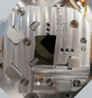
Mount samples in the dedicated Raith holder as shown in the picture, using the 3 clips available. Ensure that your sample is properly leveled, tightly kept in place by the clips, and is not obstructing the Faraday cup aperture. If the sample is insulating, you can use some conductive tape around exposure area to reduce charging.
Vent and open the SEM chamber, insert the holder on the LEO stage dovetail, close the chamber and pump down as usual.
Basic exposure
Set microscope main parameters
Once the vacuum is ready, start operating the microscope as in regular imaging. Be careful on where you are with your stage when activating the EHT, and always pay attention during stage navigation if the beam is not blanked. Any area which is imaged, is also going to be exposed, so you may risk destroying your pattern.
Set acceleration voltage, aperture and working distance according to your recipe.
Beam shaping
Find a suitable spot to optimize the beam. The quality of your pattern is directly related to the spot size of your beam and aberrations and misalignment, such as astigmatism or wobbling, will negatively affect your final resolution as well.
Since imaging through a uniform resist is not easy, you can scratch a corner of your chip to remove the resist and have some features to image. Some users pattern metal index marks on their sample surface to help navigation, which can be used to help beam shaping as well. To help with the calibration step you can mount on one of the clips a suitable dummy chip, such as an uncoated substrate or something with sub-micrometric grains, dots or lines and perform the core optimization there.
Correct stigmation and wobbling until you are satisfied with the result. Try to have good image quality at 100K or greater magnification. As long as beam parameters are left unchanged afterwards, only minor adjustments should be needed when navigating in different spots around your sample.
Measure current
Navigate with your stage until you can see the Faraday cup in the SEM image. Centre your image on the aperture and zoom all the way in. Select Patterning -> Beam Current -> Measure. Remcon32 will show a bunch of lines of code and the current will be returned after a few seconds.
You can repeat this procedure immediately or at a later time to check that the current is stable.
Chip alignment
In order to move beam and stage automatically, it is necessary to transform the software reference system (U-V) into the stage navigation one (X-Y) and vice versa. This process corrects the rotation between the two systems, and it is performed by providing two points on your sample's horizontal axis, one being the origin. At any time, you can see where you are in the UV or XY space by looking at the coordinates in the bottom right corner of Elphy.
Move to your sample. Find its bottom left corner - use the crosshairs to ensure your corner is centered in your image. Go to the Adjustments tab in Elphy, and in the Adjust UVW section select Origin Correction and click on Adjust. The UV coordinates in the bottom right corner should now zeroed. This is now the origin (0;0) point of the internal UV system. Be sure that the tab is displaying "Adjust UVW (Global)": if not, click on the Global button and repeat the adjustment.
In the same section, move to Angle Correction. Click on the P1 pipette to set the origin point as P1. Navigate to the lower right corner and do the same for P2 at that point. The tilt angle with respect to the image will be computed: click on Adjust and it will turn green. Your P1-P2 line is now the horizontal (U) axis of the internal coordinate system.
Write down the new updated coordinates of the lower right corner. It should be some form of (N;0). Do the same for the upper corners. You now know which boundaries you have available to perform your patterning. Try to avoid exposing too close to the chip edges, to avoid destroying your patterns when using tweezers or clips to hold your samples.
Set writing field
The writing field is the biggest area which the beam can expose without moving the stage - your GDS file must not exceed your selected writing field dimensions. Patterning a bigger area is typically performed by breaking your layout in an array of smaller GDS files with a suitable size. However, due to a distinct lack of accuracy in LEO stage movements (as bad as +/- 5-10 µm), stitching is highly discouraged. If your layout is too big, scale up the writing field instead.
The writing field is mainly dependent by working distance and magnification. To see, set and modify writing fields, click on ???-???-???. You can modify them by double clicking. A default working configuration is provided during training.
Once you have a suitable configuration, right click on it and click "Set"???. If everything worked, you should see the corresponding magnification will be updated on the microscope screen. You can zoom in and out while imaging: magnification will be reset to the right value when starting with the exposure.
Exposure parameters
In order to set the basic exposure parameters, go to the exposure tab and click on ???. Disable line, dot and curved elements.
Exposure parameters are connected by the following equation (visible in the pop-up window???), so they can't all be set at will at the same time. Also the current should read as the value previously measured and is not supposed to be modified.
Eq. dose=current*dwell/stepsize*linesize???
Select "equal step size"???, then input the desired values for step size and dose. Click on the little calculator button next to dwelling time - the software will compute the closest scan speed corresponding to that configuration. Click on the calculator next to dose - the software will update this value to a close one, to compensate for the non-continuous values available for the other parameters. This is your base dose, so remember to save it somewhere.
Exposure
Perform one last time image optimization (focus, stigmation, wobbling, ...) on the microscope. Try to be as close as possible to your zone of interest, without directly exposing it.
Click on File -> New positionlist???. Open the Layout??? tab and drag and drop your pattern from the list into the positionlist. Select the layers you want to be patterned. Double click??? on the new positionlist item and modify ??? with the UV coordinates where you want your pattern to be (e.g. in the middle of your sample) - this is where your chip alignment step becomes relevant. Dose???
By clicking on advanced settings, it will show the exposure parameters for this layout. it is possible to modify the basic exposure parameters previously set by unchecking the default box??? and clicking ???. The procedure is the same as above, using the calculators to satisfy the governing equation, but the parameters modified this way won't be carried to other items in the position list.
When you're satisfied with the patterning settings, click on "Times". Elphy will fracture your layout into the elementary paths which will use to move the beam when performing the exposure. If everything was done right, it will end up showing the total time which will be needed for your exposure.
Close the pop-up??? and look back at the position list. It is possible to see and modify the name of the GDS files, its UV coordinates for patterning, which layers were selected, and other useful parameters such as the dose factor (the whole pattern's dose will be multiplied by this factor, useful e.g. for a dose test). This is the last moment to spot critical errors: double check that everything is as intended.
When you are ready, highlight the item you want to expose and click the play icon???, or press F9. The software will repeat the same routine seen when clicking on "Times", but this time afterwards it will start the actual patterning, being busy for the duration computed before. Don't use the SEM or Raith computer during this time to avoid crashes or errors. When the patterning is finished, the light next to it in the position list will turn from blue to green. If it is red, something went wrong.
It is possible to add multiple elements to the position list, and start it only afterwards, by highlighting them all. This is especially useful for small fast exposures, e.g. in case of an array of a same pattern with different doses. For longer sessions, or if the stage is moved significantly, it is however advisable to move back to a corner to repeat the alignment step, since drifts in the instrument may deteriorate your later results.
After each exposure is over, the beam will be off and the stage will be right above the working area. If you need to do something else, do not turn it on there or you will destroy your pattern! Navigate to a safe spot (e.g. the one corner at U,V = 0,0) and turn on the beam only there by clicking on the beam icon and the PAT icon in the toolbar???
Unload and Shut off
Once everything is set and done, vent the chamber and unload your sample. Close Elphy on the Raith PC and Remcon32 on the LEO pc. Terminate your SEM LEO session as usual. The Raith PC remains operational even when LEO is locked.
Remember to place the Raith holder back in its box, and when logging out on Labmanager to fill the log book with "Raith session" = "Yes" and any relevant comment.
Troubleshooting
The mouse/keyboard don't work
The microscope mouse and keyboard are normally set to move freely between the LEO and the Raith screens. If this is not working, be sure that the "Scroll Lock" key on the keyboard is not active. If it is, deactivate it and it should work again. If it still doesn't work, it is possible that a Windows pop-up requires some input (e.g. Admin permission), or the computer needs rebooting. In both cases, a second set of keyboard and mouse is available on the right of the table, which is connected directly (and only) to the Raith pc.
Elphy took forever to load and then gave me an error.
Double check that Remcon32 is active on the LEO pc, and that the port COM 6 has been opened (Right click in the terminal -> Open Port).
Software doesn't perform exposure
Before starting an exposure, it may be necessary to clear error messages given by the system. Common error sources are a closed Remcon32 port, an exposure canceled after starting or one failed. Double click on the error bar in the bottom right corner and check that error messages correspond to problems already solved. Select Clear Messages and the error bar should turn green and indicate OK. If the error bar doesn't clear, check the error message and try to address the issue. If it can't be solved (or is already solved but still won't clear), consider rebooting Elphy.
If the error bar is already green but the exposure is still not performed, select the first item on the position list go to Properties, Advanced Settings and click Times, and check that the exposure can actually be completed. Incorrect parameter settings may be present, such as an unsolvable dose/dwell time/step size combination selected, or the wrong writing field.
The LEO SEM image is all black
Check that the EHT is on and everything is ok on the microscope side.
Check that the beam icon in the Elphy top bar is not blanked (beam with a red cross on it). You can click on it anytime to toggle the beam on and off.
Check that imaging mode in the Elphy top bar is active (grey "IMG" letters on white). You can click on that button to toggle between imaging and patterning (white "PAT" on blue) mode. When exposing, patterning mode should activate automatically.
The LEO SEM image is not all black, I'm exposing my resist everywhere!
If you are trying to navigate the stage automatically, check that the Auto beam off checkbox in the Stage control tab is checked.
Sometimes when starting an exposure, the software misbehaves and resumes the full scanning, exposing the whole writing field. If it happens, stop immediately the exposure (STOP button) and blank the beam manually.
If your resist has a high dose to clear (or the beam current is low) it may still be ok to try patterning that area, but it is highly recommended to just consider that spot completely exposed and discard it.
Current is too low/too high/not read/not stable
Although the current in the LEO SEM is not constant, it should not vary more than some 10-20% between different sessions and be fairly stable during each session.
If the Faraday cup reading is significantly different than what regularly experienced, double check that the image is properly centered on the Faraday cup aperture, that the maximum magnification is selected (everything should be black), and especially that the correct aperture, EHV and working distance are currently selected within the microscope.
Gun issues may reflect into the beam current stability. If you believe the current readings are no more as they should, feel free to contact a Nanolab responsible.
The stage coordinates (or other parameters) on the microscope are not updated into the Raith software.
Toggle the lightbulb button ON on the top navigation bar in order to start the continuous polling of the SEM parameters.
I moved to some known coordinates/stored position but it's not where it should be
Check that stage rotation and tilt have the same value as when the position was saved. The software stores only X, Y values, not R, T or Z.
Check that you are using the right set of coordinates (UV versus XY).
If using UV coordinates, be sure that no modification has been performed after noting those coordinates, such as adjusting a new 0 coordinate.
If using XY coordinates, the stage may not be initialised properly. Perform a stage initialisation routine.
Obsolete
Pepare beam for writing
Before you start preparing your beam, a few recommendations:
- It is recommended to move the stage (by joystick) instead of deflecting the beam (ctrl + tab); this to ensure that you work with an undeflected beam while preparing the beam for patterning and while patterning your GDS.
- It is recommended not to rotate the scan by the 'scan' knob; this...
- Measure beam current:
- Click 'Stage Control/Positions/Faraday's cup', make sure the stage moves to the center of the Faraday's cup, increase the magnification to 100.000x or more
- Toggle beam blanker to switch on beam
- Make sure you operate at a working distance of 5 mm
- Click 'XX' to measure beam current
- Move the stage to a corner of your chip. With the joystick (or from the 'stage' tab in the SEM software), rotate the stage to align the chip
- Adjust beam quality, i.e. focus, astigmatism, wobbling at a magnification of 100.000 x or more
- Move to a new spot on the chip and switch the SEM to 'spot mode'; burn a spot in the resist (approximately 20s). Correct astigmatism and aperture alignment on that spot.
- Burn a spot with a larger magnification and adjust beam quality again.
- Burn a spot at a lower magnification to see that the spot is circular at this magnification
Stage adjustment
XY is stage coordinate system, UV is sample coordinate system:
- Origin correction:
- Click 'Adjustment/Adjust UV/Origin correction'
- Move the stage to the lower left corner of the chip
- Enter (U,V)=(0,0) and click 'adjust'
- Angle correction:
- Click 'Adjustment/Adjust UV/Angle Correction', make sure the system is in 'Global' mode
- Turn on the crosshair (SmartSEM/View)
- Set magnification to approximately 100x
- turn off beam and click 'Label1/flash'-icon to move stage to the origin.
- Click 'label1/pippette'-icon
- Move stage to the lower right corner of the chip
- click 'label2/pipette'-icon.
- Click adjust: the button hereafter turns green
Exposing
- Click 'Design/open' to open a GDS file. Click 'View/Hierarchy/Max' from the menu bar to see full structure
- Set the writing field:
- Click 'Raith/Writefield Manager', right-click on the writing field you wish to use and click 'SET'
- Click 'File/Positionlist' and drag your GDS-file to the position list.
- Right-click on the pattern name in the positionlist and click 'properties':
- Enter center position of pattern in (U,V) coordinates
- Click 'Layer' and select layers of the pattern to expose
- Click 'Patterning Parameters', click the calculator icon.
- Select Area/Line/Dot and enter area dose/line dose/dot dose.
- Enter step size (beam shot pitch) and line spacing for area/line/dot
- Click calculator icon again, click 'OK' when 'OK' is highlighted
- Go to the positionlist, right-click on the pattern to expose and select 'Properties/patterning parameter/Times'
- Activate the positionlist.
- Click 'Scan/selection' from the menu bar. Exposure will start.
