Specific Process Knowledge/Characterization/EDX: Difference between revisions
No edit summary |
No edit summary |
||
| Line 1: | Line 1: | ||
'''Feedback to this page''': '''[mailto:labadviser@danchip.dtu.dk?Subject=Feed%20back%20from%20page%20http://labadviser.danchip.dtu.dk/index.php/Specific_Process_Knowledge/Characterization/ | '''Feedback to this page''': '''[mailto:labadviser@danchip.dtu.dk?Subject=Feed%20back%20from%20page%20http://labadviser.danchip.dtu.dk/index.php/Specific_Process_Knowledge/Characterization/EDX2 click here]''' | ||
= Energy Dispersive X-ray analysis (EDX) = | = Energy Dispersive X-ray analysis (EDX) = | ||
Revision as of 14:20, 4 May 2016
Feedback to this page: click here
Energy Dispersive X-ray analysis (EDX)
The technique of extracting information from the X-rays generated in a sample that is irradiated with electrons is called energy dispersive X-ray analysis or EDX. (Other acronyms are Energy Dispersive x-ray Spectroscopy, EDS, or Electron Probe Microanalysis, EPMA). The energetic electrons in the incident beam create core level vacancies as they collide with sample atoms electrons in a multiple scattering process. This leaves the atoms in the sample in an excited state. In the process of decaying from this state photons may be emitted. The energy of these photons is determined by the difference in energy of the shells involved. Since atomic shells are unique for every element so will be the transitions between them. Thus, every element has its own characteristic X-ray spectrum that can be used to determine the elemental composition.
Adding an EDX detector to a SEM provides a very powerful tool for elemental analysis. The capability of the SEM to precisely maneuver the electron beam in a number of ways enables us to make point-like analysis with nanometer precision.
Spatial resolution using EDX
Using the the very fine beam of electrons from a SEM one is capable of making point-like elemental analysis of the sample. A multiple scattering process will occur when the incident electrons collide with the sample electrons. This process generates low energetic secondary electrons and it continues until the incident electrons have lost so much energy that they are not different from the secondary electrons. The volume of the sample inside which this process takes place is called the interaction volume. X-rays are generated throughout the interaction volume and we are therefore probing the whole volume when using the EDX detector.
Using some empirical equations one can calculate how the distribution of X-rays generated in the sample will be at certain high voltages. Below are shown the depth profiles of X-rays from silicon and gold when irradiated with 5-30 kV electrons.
- Curves that show the depth of origin of the X-rays
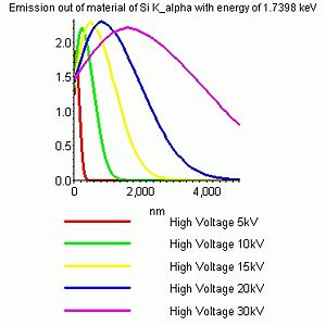
The depth profiles of X-rays emerging from bulk silicon at different high voltages.
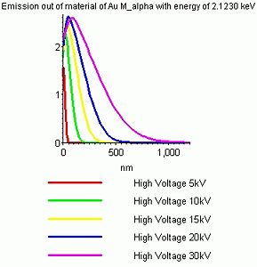
The depth profiles of X-rays emerging from bulk gold at different high voltages.


It is seen that the trend is:
- Increasing the high voltage gives rise to a bigger and deeper interaction volume and hence increases the depth which is probed.
- Increasing the density of the sample material reduces the size and depth of the interaction volume - hence a more shallow layer is probed.