LabAdviser/314/Preparation 314-307: Difference between revisions
| Line 26: | Line 26: | ||
[http://www.microscopy.uk.com/pdfs/Leica_MZ75.pdf] | [http://www.microscopy.uk.com/pdfs/Leica_MZ75.pdf] | ||
[[File: | [[File:Leica MZ75.jpg|400px|left|thumb|Location at DTU Nanolab Building 307, Room 906]] | ||
<br clear="all" /> | <br clear="all" /> | ||
==Ion milling == | ==Ion milling == | ||
Revision as of 11:26, 3 October 2019
Feedback to this page: click here
Optical Microscopes
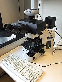
1) Olympus BX51 colorview.
2) Olympus SZ61 stereo Zoom Microscope.
3) Leica MZ75 stereo microscope.
File:Operation Manual Leica_MZ75.pdf
Ion milling
1) Fischione Low Angle Ion Milling and Polishing System Model 1010. The Fischione Ion Mill is used for producing TEM specimens with large electron transparent areas. Samples are pre-thinned by e.g. mechanical grinding and polishing or chemical polishing. The milling or polishing is performed by argon ions. The instrument has two independently adjustable Hollow Anode Discharge (HAD) argon ion sources, which can be operated from 0.5 to 6.0 kV with currents from 3 mA to 8 mA. The milling angle can be adjusted in the range of 0° to 45°. Tuning the milling parameters allows for coarse or gentle milling and the goal is to get clean samples free of physical or chemical artifacts
File:Operation Manual Fischione_1010.pdf
.
2) Fischione NanoMill model 1040. This TEM specimen preparation system is an excellent tool for creating the high-quality thin specimens needed for advanced transmission electron microscopy imaging and analysis. It is ideal for both post-FIB (focused ion beam) processing and the enhancement of conventionally prepared specimens.
Pumping stations
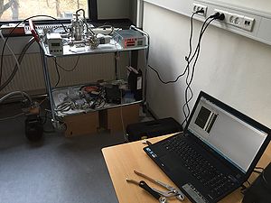
1) Pumping Cube for single TEM specimen holder.
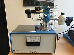
2) Gatan Turbo Pumping Station model 655. Capacity 4 specimen holders.The turbo pumping station is a completely self-contained, compact, benchtop pumping station designed to regenerate the sorb material in cooling and cryo-transfer holder dewars and for vacuum storage of transmission electron microscope (TEM) holders or samples.
Polishing
1) Struers Labopol -25.
2) TechPrep Allied.The MultiPrep™ System enables precise semiautomatic sample preparation of a wide range of materials for microscopic (optical, SEM, FIB, TEM, AFM, etc.) evaluation.
Capabilities include parallel polishing, angle polishing, site-specific polishing or any combination thereof. It provides reproducible results by eliminating inconsistencies between users, regardless of their skill.
3) Struer Labopol -5.
4) Struer Labopol -4.
Saws
1) Struers Minitom.
2) Well Precision Diamond Wire Saw.
Plasma cleaner & Pumping Station
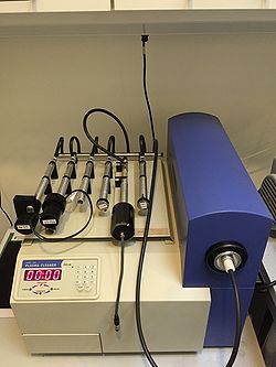
1) Fischione Plasma Cleaner model 1020. The Plasma Cleaner automatically and quickly removes organic contamination (hydrocarbon) from electron microscopy specimens and specimen holders. A low energy, reactive gas plasma cleans without changing the specimen’s elemental composition or structural characteristics. The Plasma Cleaner features easy-to-use front panel controls and an oil-free vacuum system for optimal processing.
.
Coaters & Accessories
1) Cressington 208 Carbon Coater.
2) Quorum Q150T Sputter Coater and Glow discharge
Sonicators
1) Ultrasonic Branson 2510.
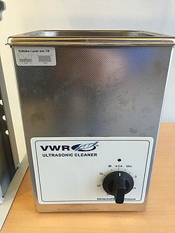
2) VWR Ultrasonic Cleaner.
3) Sonic Dismembrator.
For further information on the machine usage contact: mktracy@dtu.dk.
Hot plates
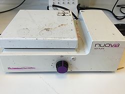
Nuova hot plate.
Plunger
Leica EM GP2
The Leica plunge freezer (EM GP2) is used to vitrifying (freezing) samples for cryo-TEM under liquid ethane temperature. For further information on the machine usage contact merafik@dtu.dk or mktracy@dtu.dk.
Microtomes
1) Ultramicrotome
RMC MT-7 Microtome
Our RMC MT-7 Microtome is a vintage piece of equipment and can be used for planning specimens for SEM/EDX and/or for cutting relative thin slices (approx. 200nm thick) for TEM. First, the specimen needs to be embedded into a resin or an epoxy, and then can be sliced with a glass or diamond knife. At DTU Nanolab Cen we can offer only glass knifes. The Risk Assessment for the RMC MT-7 Microtome can be found here File:APV RMC Microtome.pdf The RMC MT-7 Microtome is located in building 314, in the Soft Mater preparation lab. For further information on the machine usage contact: afull@dtu.dk.
Leica EM UC7
The Leica ultramicrotome is used for ultra thin sectioning of sample embedded in resin block. Glass or diamond knife is used for ultra sectioning of the samples. The Leica EM UC7 operating manual is here File:EM UC7 Operating manual.pdf and the Risk Assessment here File:Risk assessment-Ultramicrotome EM UC7.docx. For further information on the machine usage contact: mktracy@dtu.dk.
2) Cryo-Ultramicrotome
Leica EM FC7
The Leica EM FC7 operating manual is here File:EM FC7 Operating manual.pdf and the Risk Assessment here File:Risk assessment-Cryo-Ultramicrotome EM FC7.docx. For further information on the machine usage contact merafik@dtu.dk or mktracy@dtu.dk.
Critical point dryer
Leica EM CPD300
The Critical Point Drier (CPD) is used for drying samples for the SEM. The machine uses the fact that at the critical point the solvent can be converted from liquid to gas without crossing the interfaces between liquid and gas (no surface tension) and hence no sample drying artefacts. CO2 is the solvent of choice as its triple point is suitable for biological samples (31°C and 74 bars). The CO2 is not miscible with water; therefore the sample should be dissolved in acetone or ethanol. The Leica EM CPD300 operating manual is here File:EM CPD300 Operating manual.pdf and the Risk Assessment here File:Risk assessment-EM CPD300.docx.
The Leica EM CPD300 is located at DTU Nanolab Cen, building 314, in the Soft Mater (toxic) Lab. For further information on the machine usage contact mktracy@dtu.dk.
Centrifuge
The Mini Spinner Eppendorf is a desktop centrifuge with supplied rotor with maximum speed/force 13,400rpm/12,100xG. It has a 30-minute timer, which is settable in 15-sec increments. It has a short-spin mode fixed at maximum speed. The Mini Spinner Eppendorf is located in building 314, in the Soft Mater (toxic) Lab. For further information on the machine usage contact: mktracy@dtu.dk.
pH meter
If you are planning on doing chemical fixation on biological samples you will probably need to make buffers and use a pH-meter. The pH-meter (model pH 1000L) is located in building 314, in the Soft Mater (toxic) Lab and it is dedicated to “Soft Matter” related work. For further information on the machine usage contact: mktracy@dtu.dk.
