LabAdviser/314/Preparation 314-307/Soft-matter
THIS PAGE IS UNDER CONSTRUCTION
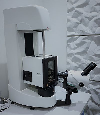
Plunge Freezer
Leica EM GP2
The plunge freezing is a cryo-fixation method used to preserve samples in their most native state prior to cryo-electron microscopy.
The main steps of this technique proceed as follows:
1. The sample is spread into a thin film across an EM grid
2. The liquid droplet is then blotted with filter paper until only a very thin film of fluid remains
3. The grid is then rapidly plunged into a cryogen (usually liquid ethane)
4. The grid is afterwards stored in liquid nitrogen in grid box until finally loaded into the cryo-electron microscope for imaging
Operating manual: File:Leica EM GP2 Operating manual.pdf
Risk Assessment: File:Risk assessment - EM GP2 Plunge Freezer.pdf
For further information on the machine usage or training contact mktracy@dtu.dk [1].

Microtomes
RMC MT-7 Microtome
Our RMC MT-7 Microtome is a vintage piece of equipment and can be used for planning specimens for SEM/EDX and/or for cutting relative thin slices (approx. 200nm thick) for TEM. First, the specimen needs to be embedded into a resin or an epoxy, and then can be sliced with a glass or diamond knife. At DTU Nanolab Cen we can offer only glass knifes. We also offer the possibility to make glass knives using the LKB Knifemaker 7801A.
Risk Assessment of RMC MT-7 Microtome: File:APV RMC Microtome.pdf
Manual of LKB Knifemaker 7801A: File:LKB Knifemaker 7800B Manual.pdf
For further information about the machine usage contact afull@dtu.dk [2].

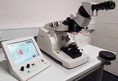
Leica EM UC7
The Leica ultramicrotome is used for ultra thin sectioning of sample embedded in resin block with a feed range of 1 nm up to 15 µm. Glass or diamond knife is used for the ultra sectioning of the samples.
Operating manual: File:EM UC7 Operating manual.pdf
Risk Assessment: File:Risk assessment - EM UC7 Ultramicrotome.pdf
For further information about the machine usage or training contact mktracy@dtu.dk [3].

Procedure of ultra-sectionning in images:


Cryo-Ultramicrotome Leica EM FC7
Operating manual: File:EM FC7 Operating manual.pdf
Risk Assessment: File:Risk assessment - EM FC7 Cryo-Ultramicrotome.pdf
For further information about the machine usage or training contact mktracy@dtu.dk [4].

Critical point dryer
Leica EM CPD300
The Critical Point Drier (CPD) is used for drying samples for the SEM. The machine uses the fact that at the critical point the solvent can be converted from liquid to gas without crossing the interfaces between liquid and gas (no surface tension) and hence no sample drying artefacts. CO2 is the solvent of choice as its triple point is suitable for biological samples (31°C and 74 bars). The CO2 is not miscible with water; therefore the sample should be dissolved in acetone or ethanol.
Operating manual: File:EM CPD300 Operating manual.pdf
Risk Assessment: File:Risk assessment - EM CPD300 Critical Point Dryer.pdf
For further information on the machine usage or training contact mktracy@dtu.dk [5].

Centrifuge
The Mini Spinner Eppendorf is a desktop centrifuge with supplied rotor with maximum speed/force 13,400rpm/12,100xG. It has a 30-minute timer, which is settable in 15-sec increments. It has a short-spin mode fixed at maximum speed. The Mini Spinner Eppendorf is located in building 314, in the Soft Mater (toxic) Lab.
For further information on the machine usage contact mktracy@dtu.dk [6].

pH meter
If you are planning on doing chemical fixation on biological samples you will probably need to make buffers and use a pH-meter. The pH-meter (model pH 1000L) is located in building 314, in the Soft Mater (toxic) Lab and it is dedicated to “Soft Matter” related work.
For further information on the machine usage contact mktracy@dtu.dk [7].

Sample preparation procedures
For further information about the procedures contact mktracy@dtu.dk [8].
Feedback to this page: click here
